Tuesday, August 25, 2009

Aneurysmal Bone Cyst of the Fifth Metacarpal


By Selahattin Ozyurek, MD; Osman Rodop, MD; Ozkan Kose, MD; Feridun Cilli, MD; Mahir Mahirogullari, MD
ORTHOPEDICS 2009; 32:606

Abstract

Aneurysmal bone cyst is a rare, rapidly growing, and destructive benign bone tumor that even more rarely involves the bones of the hand. Various treatment options for aneurysmal bone cyst have been reported in the literature, but controversy exists regarding optimal treatment. Due to its rarity in the hand, no evidence-based treatment regimen has been established.

A 21-year-old man presented with a history of pain and local swelling over his fifth metacarpal of 5 months’ duration. Physical and radiographic examination of the hand was consistent with aneurysmal bone cyst. After biopsy, pathologic examination confirmed the diagnosis of aneurysmal bone cyst. En-block resection of the tumor and autologous bicortical strut graft fixation with Kirschner wires was performed. The hand was immobilized in a short arm cast for 3 weeks after the patient received 3 weeks of physiotherapy. Kirschner wires were removed 6 weeks postoperatively. Excellent clinical and functional results were obtained with no recurrence after 3 years of follow-up with en-block resection and reconstruction with iliac crest graft. Radiographic examination demonstrated the osseous integration of the graft with no signs of recurrence.

Although treatment should be planned individually according to lesion site and size and to patient age, we suggest en-block resection to prevent recurrence and secondary surgical interventions particularly in cases with no articular involvement.


advertisement

Aneurysmal bone cyst is a rare, rapidly growing, and destructive benign bone tumor. Jaffe and Lichtenstein1 described aneurysmal bone cyst in 1942 as a distinct pathological entity by clearly separating it from hemangiomas of the bone and from other tumors in which giant cells were also a prominent feature. It is a rare tumor and accounts for 1% to 2% of all primary bone tumors.2 Aneurysmal bone cysts usually occur in the first 2 decades of life and exhibit a slight female preponderance.2,3 Aneurysmal bone cyst shows an evident predilection for long bones and the vertebral column, particularly the femur, humerus, tibia, and fibula. However, aneurysmal bone cysts arising from long bones of the hand occur rarely. Less than 5% of all aneurysmal bone cysts involve long bones of the hand.4

The pathogenesis of aneurysmal bone cyst is obscure. Lichtenstein5 suggested that persistent local disturbance in hemodynamics (venous thromboses or arteriovenous aneurysm) causes marked increase in venous pressure and leads to development of a dilated engorged vascular bed. Some authors proposed that aneurysmal bone cysts arise on a preexisting bone lesion as a secondary reaction.6,7 Trauma has been implicated as an initiative factor due to the fact that aneurysmal bone cyst is preceded by trauma with fracture or subperiosteal hematoma in some cases.8 However, most authors agree that trauma draws attention to a preexisting lesion.9,10 Recently, the genetic basis of aneurysmal bone cyst has been investigated, and specific chromosomal translocations have been reported.2,11,12 Furthermore, overexpression of insulin-like growth factor 1 is postulated to play a role in the pathogenesis.2,13

The natural history of aneurysmal bone cyst has been described as evolving through 4 radiologic stages: initial, active, stabilization, and healing.14 In the initial phase, the lesion is characterized by a well-defined area of osteolysis with discrete elevation of the periosteum. This is followed by a growth phase, in which the lesion grows rapidly with progressive destruction of bone and development of the characteristic blown-out radiologic appearance. The growth phase is succeeded by a period of stabilization, in which the characteristic soap bubble appearance develops as a result of maturation of the bony shell. Diagnosis generally occurs during the active or stabilization phase. Final healing results in progressive calcification and ossification, with the lesion transformed into a dense bony mass.

Histologically, aneurysmal bone cyst is composed of cavernous or slit-like hemorrhagic spaces surrounded and traversed by fibrous septa containing spindled cells, inflammatory cells, and a lesser number of osteoclast-like multinucleated giant cells that are often distributed around the hemorrhagic, cystic spaces. Typically, osteoid formation with or without osteoblastic rimming is observed.2,15

Various options for the treatment of aneurysmal bone cyst have been reported in the literature,16 but controversy exists regarding optimal treatment. Due to its rarity in the hand, there is no established evidence-based treatment regimen. This article presents a case of aneurysmal bone cyst affecting the fifth metacarpal that was treated by en-block resection and reconstruction with bicortical iliac crest graft.

Case Report

A 21-year-old man presented with a history of pain and local swelling over his fifth metacarpal of 5 months’ duration. On physical examination, the lesion was firm and immobile and there was slight tenderness with palpation. Active range of motion of his fifth metacarpophalangeal joint was slightly restricted, and pain was aggravated with movement. There was no history of trauma. His past medical history revealed no abnormality.

Radiographic examination of the hand showed a marked increase in diameter along the fifth metacarpal and widening of the medullary canal. The cortex was uniformly thin and the metacarpal head was spared (Figure 1A). The characteristics of the lesion were consistent with aneurysmal bone cyst. After biopsy, pathologic examination confirmed the diagnosis of aneurysmal bone cyst (Figure 1B).

Figure 1A: Radiograph of the hand at admission Figure 1B: Pathologic appearence of the tumor

Figure 1: Radiograph of the hand at admission (A). Pathologic appearence of the tumor (B; hematoxylin-eosin, magnification ×40).

En-block resection of the tumor was performed through a dorsal longitudinal incision over the fifth metacarpal. The metacarpal head, together with its carpometacarpal joint capsule, was left intact. Autologous bicortical strut graft was harvested from left iliac crest. The graft was molded into its definitive shape and inserted into the created bony defect. Multiple K-wires were used for graft fixation (Figure 2). The hand was immobilized in a short arm cast for 3 weeks after the patient received 3 weeks of physiotherapy consisting of progressive active range of motion exercises. Kirschner wires were removed 6 weeks postoperatively.

At final follow-up 3 years postoperatively, the patient had gained full range of hand motion with no pain. The patient was satisfied with the functional and cosmetic results. Radiographic examination demonstrated osseous integration of the graft with no signs of recurrence (Figure 3).

Figure 2: Early postoperative radiograph of the hand Figure 3A: Radiograph of the hand at final follow-up Figure 3B: Clinical appearence of the hand at final follow-up

Figure 2: Early postoperative radiograph of the hand. Figure 3: Radiograph (A) and clinical appearence (B) of the hand at final follow-up.

Discussion

The main goals in the treatment of aneurysmal bone cyst of the hand are eradication of the lesion, prevention of recurrence, and preservation of hand function. The literature contains conflicting knowledge about the optimal treatment method.

Currently, curettage and bone grafting is the most common operative procedure used. However, recurrence rate is high after this procedure. Basarir et al17 reported that 2 of 3 cases that were initially treated with curettage and grafting recurred. Similarly, in a case series by Frassica et al,18 curettage and bone grafting in 7 cases was associated with 4 recurrences.

However, contrary reports are also found in the literature. Ropars et al19 suggested that curettage and grafting is sufficient for treatment, and aggressive methods such as cryotherapy or resection with reconstruction should only be used in case of recurrences and articular involvement. Other authors have reported parallel successful outcomes with no relapse after simple curettage and grafting.20,21 A problem with this method is that osteoclastic activity can reabsorb the graft material, depending on the aggressiveness of the lesion. Another limitation of this method is that if graft incorporation occurs, the original size of the lesion is present and can take years to remodel. Possible recurrence after insufficient primary treatment will increase the size of the defect; the tumor may reach joint structures, and consequent bone grafting must include the epiphysis and even complete joints.

Due to the high risk of recurrence after curettage and grafting alone, various forms of adjunctive therapy have been used to decrease the rate of local recurrence.16 There are 2 cases in which cryosurgery and sclerotherapy were used as an adjuvant intralesional treatment for aneurysmal bone cyst arising in the hand.22,23 These treatments are difficult to use in the small bones of the hand and may damage surrounding intact tissue and cause serious complications such as neurapraxia, postoperative fracture, burn, infection, and wound necrosis, which may happen more easily in distal lesions. Although a 3.7% local recurrence rate was reported with cryosurgery, there is a potential risk of amputation of small bones.22,24

En-block resection and reconstruction with strut grafting is another operative treatment option. Given the aggressive nature of aneurysmal bone cysts with the tendency to develop local recurrence, en-block resection seems to be the therapy of choice. No recurrences have been reported after en-block resection in the relevant literature.10,17,18,25-29 Despite it being a curative method of treatment, its use is limited, particularly in cases where the lesion is close to articular surfaces. Articular surface reconstruction and preservation of hand function need further advanced operative techniques such as nonvascularized or vascularized toe phalanx transplantation.23,30,31 Long operation time, the need for microsurgical skills, and donor site complications are major problems associated with these techniques. Otherwise, reconstruction can only be achieved with arthrodesis, which may impair hand function.

In our case, excellent clinical and functional results were obtained with en-block resection and reconstruction with iliac crest graft with no recurrence after 3-year follow-up. The metacarpal head was spared; therefore, articular surface was left intact.

References

  1. Jaffe HL, Lichtenstein L. Solitary unicameral cyst with emphasis on the roentgen picture, the pathologic appearance and the pathogenesis. Arch Surg. 1942; (44):1004-1025.
  2. Mendenhall WM, Zlotecki RA, Gibbs CP, Reith JD, Scarborough MT, Mendenhall NP. Aneurysmal bone cyst. Am J Clin Oncol. 2006; 29(3):311-315.
  3. Leithner A, Windhager R, Lang S, Haas OA, Kainberger F, Kotz R. Aneurysmal bone cyst. A population based epidemiologic study and literature review. Clin Orthop Relat Res. 1999; (363):176-179.
  4. Campanacci M. Aneurysmal bone cyst. In: Campanacci M, ed. Bone and Soft Tissue Tumors. Bologna, Italy: Aulo Gaggi Editore; 1990:725-751.
  5. Lichtenstein L. Aneurysmal bone cyst; further observations. Cancer. 1953; 6(6):1228-1237.
  6. Biesecker JL, Marcove RC, Huvos AG, Miké V. Aneurysmal bone cysts. A clinicopathologic study of 66 cases. Cancer. 1970; 26(3):615-625.
  7. Buraczewski J, Dabska M. Pathogenesis of aneurysmal bone cyst. Relationship between the aneurysmal bone cyst and fibrous dysplasia of bone. Cancer. 1971; 28(3):597-604.
  8. Dabezies EJ, D’Ambrosia RD, Chuinard RG, Ferguson AB Jr. Aneurysmal bone cyst after fracture. A report of three cases. J Bone Joint Surg Am. 1982; 64(4):617-621.
  9. Fuhs SE, Herndon JH. Aneurysmal bone cyst involving the hand: a review and report of two cases. J Hand Surg Am. 1979; 4(2):152-159.
  10. Burkhalter WE, Schroeder FC, Eversmann WW Jr. Aneurysmal bone cysts occurring in the metacarpals: a report of three cases. J Hand Surg Am. 1978; 3(6):579-584.
  11. Panoutsakopoulos G, Pandis N, Kyriazoglou I, Gustafson P, Mertens F, Mandahl N. Recurrent t(16;17)(q22;p13) in aneurysmal bone cysts. Genes Chromosomes Cancer. 1999; 26(3):265-266.
  12. Oliveira AM, Hsi BL, Weremowicz S, et al. USP6 (Tre2) fusion oncogenes in aneurysmal bone cyst. Cancer Res. 2004; 64(6):1920-1923.
  13. Leithner A, Lang S, Windhager R, et al. Expression of insulin-like growth factor-I (IGF-I) in aneurysmal bone cyst. Mod Pathol. 2001; 14(11):1100-1104.
  14. Dabska M, Buraczewski J. Aneurysmal bone cyst. Pathology, clinical course and radiologic appearances. Cancer. 1969; 23(2):371-389.
  15. Martinez V, Sissons HA. Aneurysmal bone cyst. A review of 123 cases including primary lesions and those secondary to other bone pathology. Cancer. 1988; 61(11):2291-2304.
  16. Cottalorda J, Bourelle S. Modern concepts of primary aneurysmal bone cyst. Arch Orthop Trauma Surg. 2007; 127(2):105-114.
  17. Basarir K, Saglik Y, Yildiz Y, Tezen E. Aneurysmal bone cyst of the hand: a report of four cases. Hand Surg. 2006; 11(1-2):35-41.
  18. Frassica FJ, Amadio PC, Wold LE, Beabout JW. Aneurysmal bone cyst: clinicopathologic features and treatment of ten cases involving the hand. J Hand Surg Am. 1988; 13(5):676-683.
  19. Ropars M, Kaila R, Briggs T, Cannon S. Aneurysmal bone cysts of the metacarpals and phalanges of the hand. A 6 case series and literature review [in French]. Chir Main. 2007; 26(4-5):214-217.
  20. Sakka SA, Lock M. Aneurysmal bone cyst of the terminal phalanx of the thumb in a child. Arch Orthop Trauma Surg. 1997; 116(1-2):119-120.
  21. Sproule JA, Salmo E, Mortimer G, O’Sullivan M. Aneursymal bone cyst of the proximal phalanx of the thumb in a child. Hand Surg. 2002; 7(1):147-150.
  22. Athanasian EA, McCormack RR. Recurrent aneurysmal bone cyst of the proximal phalanx treated with cryosurgery: a case report. J Hand Surg Am. 1999; 24(2):405-412.
  23. Salon A, Rémi J, Brunelle F, Drapé JL, Glorion Ch. Total replacement of a middle phalanx by free non-vascularized chondral graft, after failure of sclerotherapy for treatment of an aneurysmal bone cyst [in French]. Chir Main. 2005; 24(3-4):187-192.
  24. Schreuder HW, Veth RP, Pruszczynski M, Lemmens JA, Koops HS, Molenaar WM. Aneurysmal bone cysts treated by curettage, cryotherapy and bone grafting. J Bone Joint Surg Br. 1997; 79(1):20-25.
  25. Gundes H, Tosun B, Muezzinoglu B, Tosun A. Total destruction of the fourth metacarpal bone by aneurysmal bone cyst: reconstruction with strut fibular graft—a case report. Hand Surg. 2005; 10(2-3):265-269.
  26. Ertem K, Karadag N, Altinok T, Karakas H. Aneurysmatic bone cyst of the second metacarpal: en-block resection and bicortical iliac crest graft replacement. Eur J Orthop Surg Traumatol. 2007; 17(1):89-91.
  27. Braatz F, Popken F, Bertram Ch, Rütt J, Hackenbroch MH. Aneurysmal bone cyst of the fourth metacarpal bone—a case report [in German]. Handchir Mikrochir Plast Chir. 2002; 34(2):128-132.
  28. Mortensen NH, Kuur E. Aneurysmal bone cyst of the proximal phalanx. J Hand Surg Br. 1990; 15(4):482-483.
  29. Kotwal PP, Jayaswal A, Singh MK, Dave PK. Aneurysmal bone cyst in the metacarpal of a child: a case report. J Hand Surg Br. 1988; 13(4):479-480.
  30. Gudemez E, Eksioglu F. Aneurysmal bone cyst of the thumb metacarpal: en-block resection and free toe phalanx transplantation. Orthopedics. 2003; 26(12):1229-1230.
  31. Rao GS, Keogh P, Webster H, Lunn PG, Burke FD. Aneurysmal bone cysts in the hand treated by free non-vascular transfer of metatarsal or proximal phalanx from the foot. J Hand Surg Br. 1993; 18(6):736-741.

Authors

Dr Ozyurek is from the Department of Orthopedics, Izmir Military Hospital, Izmir, Drs Rodop, Cilli, and Mahirogullari are from GATA Haydarpasa Training Hospital, Istanbul, and Dr Kose is from Diyarbakir Education and Research Hospital, Diyarbakir, Turkey.

Drs Ozyurek, Rodop, Kose, Cilli, and Mahirogullari have no relevant financial relationships to disclose.

Correspondence should be addressed to: Selahattin Ozyurek, MD, Department of Orthopedics, Izmir Military Hospital, Inonu Caddesi, Hatay, Izmir, Turkey.

DOI: 10.3928/01477447-20090624-25

Atraumatic Bilateral Femur Fracture in Long-Term Bisphosphonate Use


By Maria S. Goddard, MD; Kristoff R. Reid, MD; James C. Johnston, MD; Harpal S. Khanuja, MD
ORTHOPEDICS 2009; 32:607

Abstract

Postmenopausal women with osteoporosis are commonly treated with the bisphosphonate class of medications, one of the most frequently prescribed medications in the United States. In the past 4 years, reports have been published implying that long-term bisphosphonate therapy could be linked to atraumatic femoral diaphyseal fractures.

This article presents a case of a 67-year-old woman who presented with an atraumatic right femur fracture. She had a medical history notable for use of the bisphosphonate alendronate for 16 years before being switched to ibandronate for 1 year before presentation. She had sustained a similar fracture on the contralateral side 3 years previously.

This case report, in addition to a review of the literature, shows that use of the bisphosphonate class of medications for an extended period of time may result in an increased susceptibility to atraumatic femoral diaphyseal fractures. Some studies have suggested that the reason may be the mechanism of action of bisphosphonates, resulting in decreased bone turnover and remodeling. Studies have not shown if the entire class of medications produce a similar result, but patients who have been treated with any bisphosphonate for an extended period of time should be considered at risk. In patients who have already sustained a femoral diaphyseal fracture, imaging of the contralateral side should be performed to identify cortical thickening as an early sign of fracture risk. Patients should also be questioned about thigh pain.


advertisement
Cipka

Postmenopausal women with osteoporosis are commonly treated with the bisphosphonate class of medications. In 2006, approximately 22 million prescriptions for this medication were written in the United States (National Prescription Audit Plus 2006; IMS Health, Norwalk, Connecticut). However, since 2005, reports have been published indicating that long-term bisphosphonate therapy could be linked to atraumatic low-energy femoral shaft fractures.1-7 This article presents a case of a 67-year-old woman with a history of sequential bilateral atraumatic femur fractures after long-term use of bisphosphonates.

Case Report

A 67-year-old woman presented to the emergency department with a spontaneous right femoral diaphyseal fracture. There was no history of trauma and she denied antecedent pain. A review of her medical history revealed that she had sustained a left femoral diaphyseal fracture under similar circumstances 3 years previously, which had been treated with intramedullary nailing. In that earlier injury, she reported a feeling of “giving way” before falling. She had no risk factors for pathologic fractures. She was a nonsmoker and did not take corticosteroids. She had a distant history of hormone replacement therapy before 1985 to prevent osteoporosis. She began taking alendronate 70 mg per week in 1991 for osteopenia and was switched to ibandronate 150 mg monthly in 2007 for a more convenient dosing regimen. In addition, her past medical history included degenerative thoracolumbar scoliosis.

Radiographs of the pelvis, right hip, and right femur revealed a displaced diaphyseal fracture at the proximal and middle one-third right femoral junction. There was evidence of osteopenia in the metaphyseal regions (Figure 1). Given her previous history of fracture on the contralateral side and a lack of risk factors for spontaneous fractures, a magnetic resonance imaging scan without contrast was obtained to exclude a pathologic fracture. It showed no evidence of a bone lesion or other abnormality. A review of the records from her previous fracture showed no radiographic evidence of an underlying malignancy to explain that injury.

On the day of admission, she underwent cephalomedullary rod fixation. There were no postoperative complications, and she was discharged after 5 days. Pathologic analysis of bone fragments removed during the procedure showed bony trabeculae and hyaline cartilage with no evidence of granulomas or tumors.

At 6-month follow-up, the patient reported minimal pain at the fracture site. She was ambulating with a cane and maintained full range of motion. Radiographs showed the intramedullary rod in place with evidence of callus formation and a visible fracture line (Figure 2). Because of signs of delayed union, dynamization was performed by removing the distal interlocking screw. She has elected to discontinue use of any type of bisphosphonate medication since her second fall. Conventional radiographs at 1-year follow-up showed that the fracture had healed completely (Figure 3).

Figure 1: The characteristic fracture pattern of cortical thickening and a unicortical beak Figure 2A: Callus formation and visible fracture line
Figure 2B: Callus formation and visible fracture line Figure 3: A completely healed fracture

Figure 1: Preoperative conventional AP radiograph showing the characteristic fracture pattern of cortical thickening and a unicortical beak. Figure 2: Postoperative conventional AP (A) and lateral (B) radiographs taken 4 months after injury, showing callus formation and visible fracture line. Figure 3: Postoperative radiograph at 1-year follow-up visit showing a completely healed fracture.

Discussion

Some studies have suggested that long-standing bisphosphonate use may be a risk factor for atraumatic femoral shaft fractures.1-8 It is believed that bisphosphonates inhibit the normal bone remodeling cycle, thus limiting native repair and leading to the accumulation of microfractures, which places a patient at an increased risk for long-bone fractures.8,9 In a dual-center study, Odvina et al8 examined the bone biopsy results from 9 patients treated with alendronate for 3 to 8 years for osteoporosis or osteopenia; those authors proposed that the mechanism behind this paradoxical increased incidence of femoral shaft fractures is severe suppression of bone turnover by bisphosphonates. They confirmed this theory histologically as a decrease in the osteoclastic and osteoblastic surfaces and identified a reduction or lack of tetracycline labeling, indicating diminished mineralized bone.8 All patients, including those who were given estrogen therapy, had decreased bone formation and no double-tetracycline labeling.

Because bisphosphonates bind to bone and are slowly released during bone resorption,10 it may take several years for any detrimental effect to become evident. For example, alendronate has a half-life of 10.9 years,11 and therefore could be present in the body long after therapy is stopped. One study has shown that, after taking alendronate for 5 years, the biochemical markers of bone turnover remained suppressed for at least 3 years after its discontinuation.12 Using a pharmacokinetic model with a dose of 10 mg per day, Rodan et al13 found that the amount of alendronate retained in bone after 10 years of treatment was approximately 75 mg per 2 kg of mineral.

Our patient, who sustained sequential bilateral femoral diaphyseal fractures within a 3-year period, had been treated with bisphosphonates (alendronate and ibandronate) for osteopenia for more than 16 years. Given similar mechanisms without substantial trauma and no other risk factors for these fractures, the long-term use of bisphosphonates is implicated.

Alendronate has been the bisphosphonate most commonly used and has been implicated in most of the reported cases of atraumatic femur fractures. It is likely a class effect, that is, an effect related to bisphosphonates in general rather than to a specific medication. It is likely that other bisphosphonates will result in more complications as their use increases. Using a rat model, Yang et al14 showed that high levels of the bisphosphonate pamidronate lowered the bone mineral density and mechanical strength of the femur. In addition, they also found that there was reduced healing and callus formation after fracture in femurs with a high intraosseous concentration of pamidronate. They suggested that severe suppression of bone turnover also occurs with bisphosphonates other than alendronate, in high concentrations and over time.

Ott15 recommended that treatment with bisphosphonates be stopped after 5 years and that patients that require additional fracture protection be given parathyroid hormone. The rationale for this timeframe is to allow adequate time for fracture prevention while minimizing the risks of severe suppression bone turnover. A more recent study by Sebba16 suggested a medication holiday of 1 year to reduce the fracture risk from long-term uninterrupted use because there is no reduction of the protective benefit during that time. Some studies have shown that discontinuation of alendronate after this time period does not diminish the protective effect for vertebral fractures.10 It is important to define the minimal duration of treatment needed for osteoporosis to reduce the side effects of these medications.

One study has shown that treatment with estrogen replacement in combination with bisphosphonates resulted in greater levels of suppression of bone turnover than use of the latter alone.17 Although our patient had a distant history of estrogen use, it was not concomitant with her bisphosphonate therapy.

Atraumatic femoral diaphyseal fractures occurring in long-term bisphosphonate use have similar characteristics: a simple transverse pattern, unicortical beak, and cortical hypertrophy.3-6 The fracture pattern in our patient was similar to that described in other studies3-6; it appears to be pathognomonic for a femoral fracture in long-term bisphosphonate use. In a case series of 17 patients on alendronate therapy with subtrochanteric insufficiency fractures, Kwek et al3 found that all patients experienced prodromal pain and that all had similar radiographic fracture patterns. These patterns included a transverse fracture with lateral pattern, which they described as simple with thick cortices. In a 5-year retrospective review of 70 patients with low-energy femoral fractures, Neviaser et al5 found that of 25 patients being treated with alendronate, 19 (76%) had a simple, transverse fracture with a unicortical beak in an area of cortical hypertrophy. Only 1 patient of the remaining 45 who were not treated with alendronate in this study had these radiographic findings.

To our knowledge, a bilateral fracture in association with bisphosphonate use over an extended period of time has been reported in only 1 other case.1 In that study, Cheung et al1 showed suppressed bone turnover in their patient by using a double-tetracycline-labeled bone biopsy of the anterior superior iliac spine. Goh et al2 examined 9 patients who sustained subtrochanteric insufficiency fractures while on alendronate and found hypertrophy of the cortex on the contralateral side in 3 patients, implying a risk for bilateral fracture development.

Since our patient’s follow-up, we have identified 2 other patients with atraumatic fractures after bisphosphonate therapy for several years, 1 of whom had evidence of a cortical stress reaction on the opposite side. For patients presenting with a low-energy subtrochanteric or diaphyseal femur fracture and a history of long-term bisphosphonate treatment, we recommend that such medications be considered a part of the underlying abnormality. Attention should also be paid to patients who have been treated with bisphosphonates for a long time who report thigh pain because this symptom might be an early indication of an impending fracture. We also recommend that patients on bisphosphonate therapy who have already had a femoral fracture should undergo one-time imaging of the contralateral side to identify any cortical thickening. If thickening is identified, consideration should be given to a medication holiday or termination of the bisphosphonate therapy. This decision should be made in conjunction with the physician who prescribed the medication.

It seems clear that in certain patients, chronic use of bisphosphonates predisposes them to low-energy or atraumatic long bone fractures. Undoubtedly, these medications are beneficial for the prevention of vertebral compression and other osteoporotic fractures,18,19 and the discontinuation of bisphosphonates should be discussed with the patient’s primary physician. Additional pathophysiology studies are needed to identify patients who are at risk for this major complication.

References

  1. Cheung RKH, Leung KK, Lee KC, Chow TC. Sequential non-traumatic femoral shaft fractures in a patient on long-term alendronate. Hong Kong Med J. 2007; 13(6):485-489.
  2. Goh SK, Yang KY, Koh JSB, et al. Subtrochanteric insufficiency fractures in patients on alendronate therapy. A caution. J Bone Joint Surg Br. 2007; 89(3):349-353.
  3. Kwek EBK, Goh SK, Koh JSB, Png MA, Howe TS. An emerging pattern of subtrochanteric stress fractures: a long-term complication of alendronate therapy? Injury. 2008; 39(2):224-231.
  4. Lenart BA, Lorich DG, Lane JM. Atypical fractures of the femoral diaphysis in postmenopausal women taking alendronate. N Engl J Med. 2008; 358(12):1304-1306.
  5. Neviaser AS, Lane JM, Lenart BA, Edobor-Osula F, Lorich DG. Low-energy femoral shaft fractures associated with alendronate use. J Orthop Trauma. 2008; 22(5):346-350.
  6. Sayed-Noor AS, Sjoden GO. Subtrochanteric displaced insufficiency fracture after long-term alendronate therapy—a case report. Acta Orthop. 2008; 79(4):565-567.
  7. Schneider JP. Should bisphosphonates be continued indefinitely? An unusual fracture in a healthy woman on long-term alendronate. Geriatrics. 2006; 61(1):31-33.
  8. Odvina CV, Zerwekh JE, Rao DS, Maalouf N, Gottschalk FA, Pak CYC. Severely suppressed bone turnover: a potential complication of alendronate therapy. J Clin Endocrinol Metab. 2005; 90(3):1294-1301.
  9. Visekruna M, Wilson D, McKiernan FE. Severely suppressed bone turnover and atypical skeletal fragility. J Clin Endocrinol Metab. 2008; 93(8):2948-2952.
  10. Black DM, Schwartz AV, Ensrud KE, et al. Effects of continuing or stopping alendronate after 5 years of treatment. The Fracture Intervention Trial Long-term Extension (FLEX): a randomized trial. JAMA. 2006; 296(24):2927-2938.
  11. Khan SA, Kanis JA, Vasikaran S, et al. Elimination and biochemical responses to intravenous alendronate in postmenopausal osteoporosis. J Bone Miner Res. 1997; 12(10):1700-1707.
  12. Ensrud KE, Barrett-Connor EL, Schwartz A, et al. Randomized trial of effect of alendronate continuation versus discontinuation in women with low BMD: results from the Fracture Intervention Trial long-term extension. J Bone Miner Res. 2004; 19(8):1259-1269.
  13. Rodan G, Reszka A, Golub E, Rizzoli R. Bone safety of long-term bisphosphonate treatment. Curr Med Res Opin. 2004; 20(8):1291-1300.
  14. Yang KH, Won JH, Yoon HK, Ryu JH, Choo KS, Kim JS. High concentrations of pamidronate in bone weaken the mechanical properties of intact femora in a rat model. Yonsei Med J. 2007; 48(4):653-658.
  15. Ott SM. Editorial: long-term safety of bisphosphonates. J Clin Endocrinol Metab. 2005; 90(3):1897-1899.
  16. Sebba A. Osteoporosis: how long should we treat? Curr Opin Endocrinol Diabetes Obes. 2008; 15(6):502-507.
  17. Bone HG, Greenspan SL, McKeever C, et al. Alendronate and estrogen effects in postmenopausal women with low bone mineral density. Alendronate/Estrogen Study Group. J Clin Endocrinol Metab. 2000; 85(2):720-726.
  18. Black DM, Cummings SR, Karpf DB, et al. Randomised trial of effect of alendronate on risk of fracture in women with existing vertebral fractures. Fracture Intervention Trial Research Group. Lancet. 1996; 348(9041):1535-1541.
  19. Bone HG, Hosking D, Devogelaer JP, et al. Ten years’ experience with alendronate for osteoporosis in postmenopausal women. N Engl J Med. 2004; 350(12):1189-1199.

Authors

Drs Goddard, Reid, Johnston, and Khanuja are from the Department of Orthopedic Surgery, The Johns Hopkins University, Baltimore, Maryland.

Drs Goddard, Reid, Johnston, and Khanuja have no relevant financial relationships to disclose.

Correspondence should be addressed to: Harpal S. Khanuja, MD, c/o Elaine P. Henze, BJ, ELS, Medical Editor and Director, Editorial Services, Department of Orthopedic Surgery, Johns Hopkins Bayview Medical Center, 4940 Eastern Ave, #A6765, Baltimore, MD 21224-2780.

DOI: 10.3928/01477447-20090624-27

Hip Replacement Surgery

Source: http://www.bananarepublican.info/Hip_Surface_Replacement.htm

Background

Hip replacement surgery has been around since the early 1960s. Sir John Charnley experimented in the early 1950s, and he used a small (22 mm) stainless steel ball on a stem in 1962 that he inserted into the femur (hip) bone to replace the femoral head (ball). He then inserted a high-density plastic socket to replace the acetabular (socket) side of the hip joint. Both were secured with a self-curing acrylic polymer known as bone cement.

Total Hip Replacements

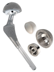
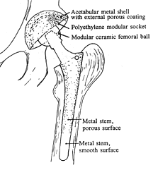
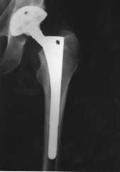
Today, the modular balls are made of a cobalt-chrome metal alloy or a ceramic material, and some of the components are press-fit and do not require bone cement. The procedure remains basically the same: (1) the femur bone is amputated to remove the femoral head; (2) the femoral canal is reamed-out for insertion of the stem; (3) an acetabular socket is affixed to the socket side of the hip; and (4) the ball joint is inserted into the acetabular socket. This is known as a total hip replacement, or more correctly, total hip arthroplasty (THA).

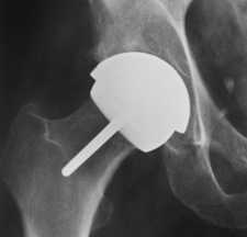
image from www.wmt.com

(Click on image to enlarge)

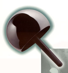
image from www.jri-oh.com

(Click on image to enlarge)

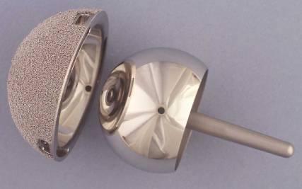
image from www.jri-oh.com

(Click on image to enlarge)

The acetabular socket used in THA is normally lined with a high molecular weight polyethylene (sometimes the liner is ceramic). A metal or ceramic ball is attached to the stem and rotates within the socket. Fine particulate debris is produced from the wearing process of the ball against the liner that leads to tissue reaction. The body’s immune system attacks the debris, and consequently, attacks the adjacent bone supporting the THA device, leading to bone loss and a loosening of the device. This bone loss is known as osteolysis. To lessen the amount of wear, a small ball (approximately 30 mm) is used; however, the small size of the ball makes the joint less stable and increases the risk of dislocation in certain circumstances.

The loosening of the THA device requires revision surgery in which a larger diameter stem must be inserted in the femoral canal. Depending on the age and activity of the patient, multiple revision surgeries may be necessary throughout a patient’s life. A young (under 60), active individual can expect only 10 – 15 years before needing revision surgery. Revision surgery can be complex and costly. The lifespan of a THA device is clocked in miles rather than years.

(Note: Wright Medical Technology, Inc. has developed a large femoral head using metal-on-metal technology (see Hip Surface Replacement below) that reduces the risk of dislocations and osteolysis in THRs. The large head THR has received FDA approval and is actively being marketed.)

Hip Surface Replacements

Although it was experimented with and attempted in the 1960s, metal-on-metal “resurfacing” of the femur and acetabulum was abandoned because of loosening of the fittings. With the refinement of acrylic fixation and its very successful use with the THA stem, interest in hip resurfacing was renewed, and it was subsequently used in several countries in the 1970s. (See History of Hip Resurfacing.)

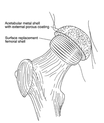
Resurfacing has the advantage of preserving the femoral bone stock (and marrow contained in the femur). It also has the advantage of easy future revision to THA if it becomes necessary. Since the femur is persevered and not amputated in the initial hip surface replacement surgery, it is available to support a THA stem should revision become necessary. Maintaining the integrity of the femur bone also aids in the mechanical transfer of weight and stress in a more natural manner. Where THA patients often experience thigh pain, recipients of hip surface replacements avoid that particular discomfort.

image from www.jri-oh.com

(Click on image to enlarge)

image from www.jri-oh.com

(Click on image to enlarge)

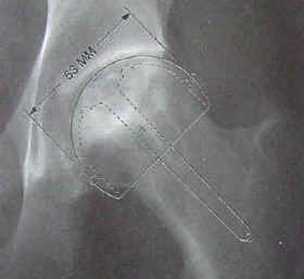
Using a metal acetabular socket as well as a metal cap over the femur head (metal-on-metal) eliminates the polyethylene debris produced in THA. The metal wear debris from a hip surface replacement produces smaller particles than polyethylene wear debris. The inflammatory response to metal debris is considerably less than that from polyethylene debris. It is believed that the body can partially dissolve and expel metal since it is a naturally occurring substance in the body. There is concern by some of the toxicity of metal, but there is currently no definitive evidence that metal ions cause cancer. Since a metal surface does not wear as readily as a polyethylene lining, a larger ball (approximately 38-51 mm) can be used that adds stability to the joint and reduces the danger of dislocation.

images from www.wmt.com & www.jri-oh.com

(Click on images to enlarge)

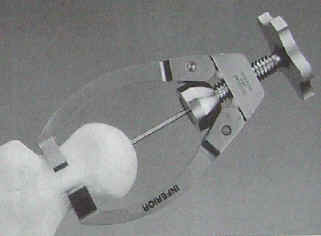
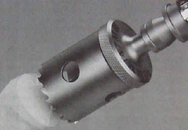
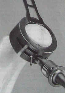
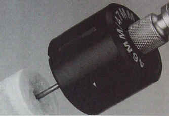
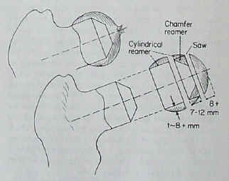
The surgery time for hip surface replacement is slightly longer than that for THA. The attachment of the acetabular socket is basically the same. It is press-fitted and does not require bone cement. The attachment of the cobalt-chrome cap requires a more precise alignment, and it takes slightly longer to fit. The hole for the pin insertion must be aligned and drilled, and the dome of the femoral head must be ground and shaped to fit the cap. Some bone cement is used to affix the cap, but the interior surface of both the cap and the socket is such that bone grows into the relief surface to grip the device. (See the video clips of hip resurfacing surgery.)

The following images are from an Instructional Lecture delivered at an International Symposium in Fukuoka, Japan on March 16, 1996 by Harlan C. Amstutz, Peter Grigoris, and Frederick J. Dorey entitled "Evolution and future of surface replacement of the hip." Journal of Orthopaedic Science. J Orthop Sci (1998) 3:169-186.

Superimposed hemisurface. Pin centering guide. Cylindrical reamer.

Saw cutoff guide and oscillating saw. Chamfered reamer. Femoral head bone preparation.

(Click on images to enlarge)

Risks involved in the hip surface replacement surgery are the same as the risks involved in any major surgery. Risks specific to the hip surface replacement involve the potential for cracking in the neck of the femur bone due to the drilling of the guide hole through the neck for the support pin in the metal cap, and also a negative reaction of the femur head to dislocation and being reshaped to fit the metal cap leading to the development of avascular necrosis (bone death)--often referred to as AVN--due to a disruption of blood circulation to the femur head and neck (see AVN Risk). In such instances, a THA could easily be performed to correct the problem.

Hip surface replacement in the United States has been pioneered by Harlan C. Amstutz, M.D. at the Joint Replacement Institute in Los Angeles, CA. For years, a hip surface replacement in the United States has been labeled an “investigative device” by the Food and Drug Administration (FDA). The longest study has been conducted by Wright Medical Technology, Inc. under the product name of CONSERVE ® Plus Total Resurfacing Hip System. The clinical trials have proceeded for a number of years, and they are nearing their end. They have involved nine surgeons across the country in California, Florida, Texas, Maryland, North Carolina, Ohio, and in the Pacific Northwest. Corin Medical, Ltd. of the United Kingdom has also begun an FDA study in the United States using the Cormet 2000 device.

Click here to read the 2-6 year follow up report of the first 400 CONSERVE ® Plus hips.

In Europe, the Birmingham Hip Surface Replacement System (BHR) has been in use for many years. Smith & Nephew Inc., manufacturer of the BHR, applied for FDA approval, and perhaps due to the long record of use in Europe, they obtained FDA premarket approval to begin commercial distribution of their device in the United States on May 9, 2006 (see FDA approval letter and FDA announcement). Because the BHR was not previously used in the United States, the number of American surgeons qualified to use it was limited due to the fact that they had all been participating in the Wright and Corin studies; however, that is destined to change with the FDA approval obtained by Smith & Nephew.

Distal Femur Defects Reconstructed With Polymethylmethacrylate and Internal Fixation Devices: A Biomechanical Study


By Anthony D. Uglialoro, MD; Michael Maceroli, BS; Kathleen S. Beebe, MD; Joseph Benevenia, MD; Francis R. Patterson, MD
ORTHOPEDICS 2009; 32:561

Abstract

Benign aggressive distal femur tumors are treated with curettage, adjuvant phenol or argon, and polymethylmethacrylate (PMMA) packing. For large defects, an internal fixation device is added to reduce the fracture risk. The purpose of this study is to compare the strength of locking plates to other fixation devices for stabilization of these defects.

Lateral condyle defects in young, fresh frozen femurs were packed with PMMA and augmented by internal fixation. Three groups of 4 matched pairs of femurs were organized for the following comparisons: (1) stacked Steinmann pins vs crossed screws; (2) stacked pins vs locking plates; and (3) crossed screws vs locking plates. Specimens were subjected to axial load-to-failure testing on an MTS machine.

There was no difference in load-to-failure strength (P=.177) using Steinmann pins or crossed screws. Locking plate constructs were stronger (P=.028) than Steinmann pin constructs. Locking plate constructs were also stronger (P<.001) than crossed-screw constructs. Steinmann pin constructs failed with severe intra-articular fractures; crossed screw constructs failed with bulging of the defects, articular impaction, and minimal fracture propagation. Locking plate constructs failed with extra-articular spiral shaft fractures.


advertisement
Cipka

The distal femur is a common site for many benign bone tumors, such as giant cell tumor of bone. Tumors of this type are typically treated via extended curettage through a large cortical window.1,2 After curettage the local bony architecture is left markedly disturbed and the host bone is vulnerable to postoperative fracture.3,4 Therefore, the lesion is typically packed with polymethylmethacrylate (PMMA) cement. Studies have shown reconstruction with PMMA has improved stability of the repaired bone while still allowing for ease of a second intervention if necessary.5,6 Despite the advantages of curettage and cementation, if a defect occupies >50% of the metaphysis and extends down to the articular surface, internal fixation is typically used to reduce the chances of postoperative fracture.7-9

In the past, various types of devices and configurations have been used to augment the PMMA in post-curettage defects, including stacked Steinmann pins and crossed screws. Studies have compared the biomechanical properties of these reconstructive techniques. In particular, Randall et al10 showed tibial defects reconstructed with Steinmann pins augmenting PMMA displayed significantly greater load to failure and survival when compared to specimens repaired with PMMA alone. Despite these results, other studies have found no significant difference between PMMA and PMMA supplemented with Steinmann pins.11,12 In a pivotal study, Toy et al13 showed that distal femur defects reconstructed with crossed screws augmenting PMMA cement displayed a significant biomechanical advantage over femurs repaired with PMMA alone or PMMA reinforced with Steinmann pins. To date, no study has compared reconstruction with Steinmann pins or crossed screws to locking condylar plates, the current preferred method of internal fixation.

Locking plates are now commonly indicated for internal fixation of peri-articular distal femur fractures, especially for those with metaphyseal communition, providing a rigid, toggle-free, fixed angle construct. Furthermore, locking plates represent an effective, post-curettage internal fixation device to support acrylic cementation in treatment of giant cell tumor. Studies in cadaveric models have demonstrated the structural superiority of locking plates over an unlocked, limited contact–dynamic compression plate (LC-DCP).14 The use of locking plates in distal femur fracture has shown a reduction in fixation failure, as well as less varus collapse.15 In addition, when used for fracture fixation, the locking plate can lie above the bone surface thereby preserving vascular supply of bone and preventing postoperative fractures, a common side effect of friction in LC-DCP reconstruction.14

Despite clinical support for the use of locking plates in bone reconstruction, to date no current biomechanical study has compared the stability of locking plates to Steinmann pins and crossed screws for post-curettage augmentation of PMMA. Our aim in this study is to determine which combination of polymethylmethacrylate (PMMA) and internal fixation device will yield the most biomechanically stable construct.

Materials and Methods

Twelve pairs of matched femora from human cadavera obtained from the Musculoskeletal Transplant Foundation (Edison, New Jersey) were divided into 3 groups each containing 4 pairs of fresh-frozen femora; a total of 8 specimens per testing group. The average age of donors at the time of death was 38 years (range, 21-54 years). The differences in age and gender were accounted for by comparing the post-fixation strength and stiffness of a given specimen to that of its matched counterpart that was internally stabilized with a different experimental fixation device. This allowed the assumption that although the mechanical resistance may be dissimilar if comparing specimens from 2 different donors, the strength and stiffness of a matched pair of femora from a single donor should be nearly identical.

Preparation of Defects

The fresh-frozen femora were thawed at room temperature. The femora were stripped of all soft tissues and radiographs were made to confirm osseous integrity. A high-speed burr (Medtronics, Fort Worth, Texas) was then used to create a defect in the lateral condyle of all specimens, extending from the junction of the metaphyseal and diaphyseal areas down to the subchondral bone. This defect represents the cavity that remains after a giant cell tumor is excised via curettage from the lateral femoral condyle. In the coronal/frontal plane, the defect extended from the edge of the lateral femoral condyle to the trochlear groove (distance x) (Figure 1). In the sagittal plane, the defect extended from the entire anteroposterior width (distance y) of the lateral femoral condyle, leaving a cortical shell intact (Figure 2). Cortical and cancellous bone was removed from the subchondral plate to the epiphyseal/metaphyseal junction (Distance x’). After the defect had been created in each matched pair, the reconstruction method was determined by a random number generator.

Figure 1: The defect extended from the edge of the lateral femoral condyle to the trochlear groove Figure 2: The defect extended from the edge of the lateral femoral condyle to the trochlear groove

Figure 1: In the coronal/frontal plane, the defect extended from the edge of the lateral femoral condyle to the trochlear groove (distance x). The vertical height was equal to this distance (x’). Figure 2: The vertical height was equal to the distance x’. In the sagittal plane, the defect extended from the edge of the lateral femoral condyle to the trochlear groove (leaving a cortical shell intact).

Methods of Fixation

Three groups of 4 matched pairs of femora were organized for the following comparisons: (1) Steinmann pins vs crossed screws; (2) Steinmann pins vs locking plates; and (3) crossed screws vs locking plates. In each respective group, 1 of the matched pairs from a single donor was randomly assigned 1 of 2 different experimental fixation devices, while its matched counterpart received another stabilization method.

For femora reconstructed with Steinmann pins, three 3/16-in threaded intra-medullary Steinmann pins (Zimmer Inc, Warsaw, Indiana) were manually inserted into the medullary canal such that they extended into the diaphysis. The distal ends of the Steinmann pins were fanned out to support the lateral joint surface (Figure 3).

Figure 3A: Post-curettage distal femur reconstructed with Steinmann pins Figure 3B: Gross image of Steinmann pin placement

Figure 3: Lateral radiograph of post-curettage distal femur reconstructed with Steinmann pins without PMMA (A). Gross image of Steinmann pin placement in the lateral femoral condyle (B).

For femora reconstructed with crossed screws (Synthes, Paoli, Pennsylvania), a 3.2-mm bit was used to drill 4 guide holes extending to the opposite cortex. Two holes were directed from the distal aspect of the femur proximally (superiorly) to the contralateral cortex, while the other 2 holes were directed inferiorly toward the opposite medial condyle. Two 4.5-mm cortical screws were placed into the superiorly directed holes, and two 6.5-mm cancellous screws were placed in the inferiorly directed holes. The screws were advanced into the bone until the screw heads were aligned with the missing border of the lateral condyle (Figure 4).

Figure 4A: The distal femur repaired with crossed screws Figure 4B: The distal femur repaired with crossed screws

Figure 4: AP (A) and lateral (B) radiographs of the distal femur repaired with crossed screws.

For femora reconstructed with locking condylar plates (Synthes) the standard, accepted method was used (Figure 5).

After all femora were reconstructed with either Steinmann pins, crossed screws, or locking plates, PMMA was prepared and mixed. The cement was molded into the defect, around the pins, screws, or locking plate and shaped to form the physiologic contour of the lateral femoral condyle. The cement was then given ample time to cure.

Figure 5A: The defect can be seen in the lateral condyle of this femur sample Figure 5B: The distal femur repaired with crossed screws Figure 6: The loading nose was centered to apply an even load to both femoral condyles

Figure 5: AP radiograph (A) and gross image (B) of reconstruction with locking plate. Note that the defect can be seen in the lateral condyle of this femur sample. Figure 6: Distal femur sample reconstructed with a locking plate shown secured in the INSTRON machine. The loading nose was centered to apply an even load to both femoral condyles.

Testing

Each femur was transected 25 cm proximal to the joint line with a hand saw. The transected femora were then placed into a stainless steel fixture and fixed into acrylic cement. All femora were set into the fixture at an angle that placed the transcondylar axis horizontally when mounted in the testing apparatus.

Each femur, set in its steel fixture, was bolted into the load frame of the Instron machine (Instron, Norwood, Massachusetts). The loading nose was centered to apply a physiological load to both femoral condyles (Figure 6). Starting at 0 load and displacement the load was increased by 10 N/sec until the load reached 475 N. The load was then cycled in a sinusoidal pattern between 50 and 900 N for 2000 cycles at 1 Hz. Each femur that survived was loaded at 1 mm/sec under displacement-controlled feedback until failure. For this trial, failure is defined as a sudden drop of 445 N from the maximum observed load (Figure 7, load vs displacement curve). Load and displacement were recorded during testing and the load vs displacement curve was plotted. From these graphs, the load to failure (N) and the stiffness (N/mm) was calculated and recorded for each femur. In addition, the mode of failure was noted by the authors, photographed, and recorded for each specimen.

Figure 7: The force vs displacement curve

Figure 7: The force vs displacement curve illustrates how we determined load to failure. A drop in Force >445 N was considered a failure. The force measurement immediately preceding this drop was determined to be the load to failure.

Statistical Analysis

Within each group, a paired t test was used to compare differences between treatments for load to failure and stiffness.

Results

All three groups of femora survived the initial sinusoidal pattern of 50 to 900 N for 2000 cycles at 1 Hz.

Group 1 consisted of femora reconstructed with PMMA and Steinmann pins versus PMMA and crossed screws. The mean load to failure for femora reconstructed with PMMA and Steinmann pins was 14916±2959 N compared to 11651±3074 N for femora repaired with crossed screws augmenting PMMA (Table 1). There was no significant difference (P=.831) in load to failure between Steinmann pin and crossed screw constructs.

Table 1: Data on Load to Failure and Stiffness

Group 2 compared femora repaired with PMMA and Steinmann pins against those fixed with PMMA and locking plates. Femora reconstructed with Steinmann pins augmenting PMMA failed at an average load of 11728±2724 N compared with 24245±8228 N for the femora reconstructed with locking plates and PMMA (Table 1). For all of the matched pairs, the femur samples reconstructed with locking plates maintained a more stable construct (P=.013) than femora repaired with Steinmann pins.

In group 3, comparing femora fixed with PMMA and crossed screws to those repaired with PMMA and locking plates, the average load to failure for femora reconstructed with crossed screws augmenting PMMA cement was 9880±1130 N while the load to failure for femora repaired with locking plates and PMMA was 22188±3622 N (Table 1). In all pairs of femora, the locking plate reconstructions were significantly stronger (P=.004) than their matched counterparts repaired with crossed screws augmenting cement. Figure 8 displays the load to failure for groups 1-3.

Figure 8: Load to failure for groups 1-3

Figure 8: Load to failure for groups 1-3. Locking plates represent a significantly stronger construct than Steinman pins and crossed screws in groups 2 and 3, respectively.

Within each matched group, the femora reconstructed with locking plates augmenting PMMA (P=.03) had significantly greater stiffness than the contralateral matched pair repaired with Steinmann pins and PMMA (Table 1). However, there was no significant difference in stiffness between locking plates versus crossed screws augmenting PMMA (P=.552). In addition, stiffness values for Group 1 femora, Steinmann pins versus crossed screws, were not significantly different (P=.334).

Table 2: Mode of Failure

Table 2 displays the mode of failure for all femur samples. All 8 femora reconstructed with Steinmann pins and PMMA failed via a severe, intra-articular (intercondylar) fracture (Figure 9). Seven of the 8 femora repaired with crossed screws augmenting PMMA showed an expanded cortex resulting in bulging and impaction of the articular surface with minimal propagation down the shaft (Figure 10). One crossed screw and PMMA construct failed via an extra-articular fracture. Failure of 6 femora reconstructed with locking plates and PMMA resulted in an extra-articular spiral fracture either anterior or posterior to the locking plate screws (Figure 11). Two of the 8 femora in the locking plate group did not show extra-articular fracture. One of these failed via a supracondylar fracture (Figure 12) while the other femur survived mechanical testing without fracture thereby reaching load capacity (~30,000 N) of the mechanical testing machine. In all femora that were fixated with LCP, the articular surface was noted to be intact without any signs of compromise or fracture.

Figure 9: All 8 femora reconstructed with Steinmann pins and PMMA failed Figure 10: Seven of the 8 femora showed bulging

Figure 9: All 8 femora reconstructed with Steinmann pins and PMMA failed via a severe, intra-articular (intercondylar) fracture. Figure 10: Seven of the 8 femora repaired with crossed screws augmenting PMMA showed bulging and impaction of the articular surface with minimal propagation down the shaft.


Figure 11: Extra-articular spiral fracture either anterior or posterior to the locking plate screws Figure 12: One locking plate reconstruction failed via a supracondylar fracture

Figure 11: Failure of 6 femora reconstructed with locking plates and PMMA resulted in an extra-articular spiral fracture either anterior or posterior to the locking plate screws. Figure 12: One of the 8 locking plate reconstructions failed via a supracondylar fracture.

Discussion

Considering the extensive curettage required to treat giant cell tumors, durable reconstruction is needed to prevent postoperative fractures. Polymethylmethacrylate cementation is accepted as the foundation for noncontained giant cell tumor defect reconstruction for both the stability and adjunctive thermal necrotic effect it offers.16 The need for reinforcement in post-curettage defects has been debated in the literature and various methods of post-curettage augmentation have been tested including stacked Steinmann pins and crossed screws. To our knowledge no studies have evaluated the biomechanical advantages offered by locking plate augmentation to PMMA for reconstruction of giant cell tumor defects.

A retrospective study conducted by Bini et al5 found that giant cell tumor patients whose lesions were fixed with threaded Steinmann pins and PMMA did not experience postoperative fractures. Randall et al10 corroborated this conclusion in a biomechanical study of lateral tibial condyles showing that defects reinforced with PMMA and Steinmann pins displayed significantly greater load to failure than those repaired with PMMA alone. More recently however, Murray et al11 found no significant biomechanical differences between femoral condyle defects repaired with PMMA and Steinmann pins or PMMA alone, further escalating the debate about the need for reinforcement in post-curettage defects.

In a biomechanical study using fresh-frozen femur samples, Toy et al13 demonstrated that distal femur defects repaired with PMMA augmented by crossed screws resulted in a stronger reconstruction than PMMA alone or PMMA with Steinmann pins. In this study, large noncontained defects were created in the medial femoral condyles of 20 matched pairs of human femurs. The femora in various groups were tested to find stiffness and load to failure, with failure being defined as a sudden drop of 445 N. In all trials, the PMMA and crossed screws group represented a significant biomechanical advantage over PMMA alone and PMMA reinforced with Steinmann pins. As noted by the authors, the use of matched pairs of femora minimized variability and allowed the focus to remain on comparing the mechanical advantages of each construct. Acknowledging the power of this study, our design was modeled closely on the methods described by Toy et al13 with the addition of PMMA reinforced with femoral locking plates.

Previously acknowledged as an effective mode of fracture fixation, the versatility of locking plates and their application in oncological treatment has been largely anecdotal. A recent study examined 25 patients receiving locking plates for oncological reconstruction. It was shown that 23 of 25 locking plates were intact after a mean follow-up of 18.2 months. These results are encouraging and show clinically, locking plates can provide a reliable and stable option for oncological reconstruction.17 Through biomechanical testing, our aim was to determine if this conclusion is applicable to distal femoral defects resultant from giant cell tumor resection and curettage. Our study was modeled to elucidate the role locking plates could play in the treatment of this benign, locally aggressive bone tumor.13

To our knowledge the present study is the first biomechanical evaluation of distal femoral locking plates used for oncological reconstruction. This study compares PMMA augmented by a distal femoral locking plate with the previously examined crossed-screw and intramedullary Steinmann pin constructs. Fresh-frozen femurs were used to best represent in vivo bone quality, and cross-matched femur pairs allowed us to control for femur quality and size providing a more powerful comparison between the constructs. Defects of the lateral femoral condyle were created to mimic those caused by giant cell tumor excision and curettage. A load-to-failure biomechanical analysis of the lateral femoral condyle was then conducted comparing cross-matched femurs reconstructed with cement and augmented with intramedullary Steinmann pins, crossed-screws or condylar locking plate.

The significant differences in load-to-failure and stiffness between the different constructs validate our hypothesis that locking plate augmented PMMA is biomechanically superior to crossed-screw or Steinmann Pin augmentation. In all pairs of femora, the locking plate reconstructions showed a significantly higher load to failure (P=.004) than their matched counterparts. The locking plates failed at forces 43% and 53% greater than the Steinmann pins and crossed-screws, respectively. Furthermore the fracture pattern differed between the groups. The Steinmann pin reinforced femurs failed through a severe intra-articular fracture. All but 1 of the crossed-screw augmented femurs failed through bulging of the articular surface (expanded cortex) and spiral fracture. One crossed screw construct failed via an extra-articular fracture. Of the femora reconstructed with plates, 6 of 8 failed via an extra-articular spiral fracture anterior or posterior to the locked screws. Of the other 2 locking plate reconstructions, 1 failed via supracondylar fracture while the other did not fail. As noted by Toy et al,13 failure via an extra-articular fracture is more desirable. These fractures often can be repaired with standard open reduction and internal fixation while fractures involving the articular surface are more difficult to salvage often requiring allograft or endoprosthetic reconstruction.

Our explanation as to why the locking plate reconstruction showed a significant advantage over Steinmann pins or crossed screws is because of the additional stiffness the plates provide. As noted by Toy et al,13 the addition of screws crossing the midline in the condylar defect improves stability of the cement mantle thus transferring the load proximally. The multiple locking screws used in the locking plate head essentially act as crossed screws used in Toy’s study. As supported by the extra-articular type fractures seen in the locking plate reinforced femora, this allows the axial compression forces applied to the condyle to be transmitted proximally to the femur shaft that has better bone quality and is undisturbed by tumor and curettage. The superior fixation provided by multiple fixed-angle cortical screws also stabilizes femoral shaft to provide additional coronal stability.

The limitations of this study need to be considered before extrapolating the results for clinical application. For biomechanical testing, the femora were mounted at an angle in the testing apparatus such that the transcondylar axis was oriented horizontally. By orienting the femora as such, the applied force mimicked physiologic load to most specifically test the strength of each construct. Perhaps future biomechanical studies could examine the effects of torsion on these 3 modes of reconstruction to further delve into the response to physiologic pressure. Additionally these results should not be extrapolated to other anatomic sites. Locking plate augmentation of PMMA also has the disadvantage of being a more technically difficult surgery than cement alone or cement augmented with crossed-screws. Furthermore the additional hardware poses greater difficulty if there is recurrence of tumor or the hardware fails and a revision surgery is required. Therefore these data must be considered as biomechanical in nature only. Although the additional strength of the construct and the clinical evidence reported by Virkus et al17 appear to support the routine use of locking plates for reconstruction of giant cell tumor defects, the aforementioned shortcomings should lead practicing surgeons to take a more selective approach in managing giant cell tumor with locking condylar plates.

In this in vitro study, reconstruction of post-curettage distal femora with locking plates and polymethylmethacrylate was biomechanically stronger in axial load testing than Steinmann pins or crossed screws augmenting PMMA. The locking plate constructs were able to withstand compression forces exceeding physiologic levels. This resilient construction may permit more rapid mobilization and prevent postoperative fracture. If the locking plate reconstruction failed, the ensuing fracture is likely to be extra-articular and would preserve the articular surface. Fractures of this type are easier to treat and rehabilitate than the intercondylar fractures associated with other modes of reconstruction. Although not recommended for routine management, locking condylar plates present a significant biomechanical advantage over opposing devices such that LCP should be the preferred reconstruction method when giant cell tumor is complicated by pathological fracture.

References

  1. McDonald DJ, Weber KL. Giant cell tumor of bone. In: Schwartz HS, ed. Orthopaedic Knowledge Update: Musculoskeletal Tumors 2 Rosemont, IL: American Academy of Orthopaedic Surgeons; 2007:133-140.
  2. McGarry SV, Gibbs CP. Giant cell tumor of bone. In Damron TA, ed. Orthopaedic Surgery Essentials: Oncology and Basic Science Philadelphia, PA: Lippincott Williams and Wilkins; 2008:159-164.
  3. Buecker PJ, Gebhardt MC. Are fibula strut allografts a reliable alternative for periarticular reconstruction after curettage for bone tumors? Clin Orthop Relat Res 2007; (461):170-174.
  4. Marcove RC, Weis LD, Vaghaiwalla MR, Pearson R. Cryosurgery in the treatment of giant cell tumors of bone: a report of 52 consecutive cases. Clin Orthop Relat Res 1978; (134):275-289.
  5. Bini SA, Gill K, Johnston JO. Giant cell tumor of bone: curettage and cement reconstruction. Clin Orthop Relat Res 1995; (321):245-250.
  6. Persson BM, Wouters HW. Curettage and acrylic fixation cementation in surgery of giant cell tumors of bone. Clin Orthop Relat Res 1976; (120):125-133.
  7. Leeson MC, Lippitt SB. Thermal aspects of the use of polymethylmethacrylate in large metaphyseal defects in bone. A clinical review and laboratory study. Clin Orthop Relat Res 1993; (295):239-245.
  8. Mjöberg B, Pettersson H, Rosenqvist R, Rydholm A. Bone cement, thermal injury and the radiolucent zone. Acta Orthop Scand 1984; 55(6):597-600.
  9. Weber KL. Surgical treatment of benign bone tumors about the knee. Techniques in Knee Surgery 2003; 2(4):274-285.
  10. Randall RL, Weenig KN, West JR, Johnston JO, Bachus KN. Durability and strength of Steinmann pin augmentation in cemented tibial defects. Clin Orthop Relat Res 2002; (397):306-314.
  11. Murray PJ, Damron TA, Green JK, Morgan HD, Werner FW. Contained femoral defects: biomechanical analysis of pin augmentation in cement. Clin Orthop Relat Res 2004; (420):251-256.
  12. Weiner M, Damron TA, Patterson FR, Werner FW, Mann KA. Biomechanical study of pins in cementing of contained proximal tibia defect. Clin Orthop Relat Res 2004; (419):232-237.
  13. Toy PC, France J, Randall L, Neel MD, Shorr RI, Heck RK. Reconstruction of noncontained distal femoral defects with polymethylmethacrylate and crossed-screw augmentation: a biomechanical study. J Bone Joint Surg Am 2006; 88(1):171-178.
  14. Haidukewych GJ. Innovations in locking plate technology. J Am Acad Orthop Surg 2004; 12(4): 205-212, 2004.
  15. Haidukewych GJ, Ricci W. Locked plating in orthopaedic trauma: A clinical update. J Am Acad Orthop Surg 2008; 16(6):347-355.
  16. Komiya S, Inoue A. Cementation in the treatment of giant cell tumor of bone. Arch Orthop Trauma Surg 1993; 112(2):51-55.
  17. Virkus WW, Miller BJ, Ping CC, Gitelis S. The use of locking plates in orthopedic oncology reconstructions. Orthopedics 2008; 31(5):1.

Authors

Drs Uglialoro, Beebe, Benevenia, and Patterson and Mr Maceroli are from the University of Medicine and Dentistry of New Jersey – New Jersey Medical School, Newark, New Jersey.

Drs Uglialoro, Beebe, Benevenia, and Patterson and Mr Maceroli have no relevant financial relationships to disclose.

The authors thank the Musculoskeletal Transplant Foundation (Edison, New Jersey) and Synthes Inc (Paoli, Pennsylvania) for sponsoring this project. The authors also thank Assimina A. Pelegri, PhD, and her staff at Rutgers University, Department of Mechanical and Aerospace Engineering (Piscataway, New Jersey).

Correspondence should be addressed to: Francis R. Patterson, MD, Department of Orthopedics, Division of Musculoskeletal Oncology, UMDNJ – New Jersey Medical School, 140 Bergen St, ACC Building, Ste D-1610, Newark, NJ 07103.

DOI: 10.3928/01477447-20090624-29

Treatment of Giant Cell Tumor of Long Bones: Clinical Outcome and Reconstructive Strategy for Lower and Upper Limbs


By Keiichi Muramatsu, MD; Koichiro Ihara, MD; Toshihiko Taguchi, MD
ORTHOPEDICS 2009; 32:491

Abstract

Giant cell tumor of bone is a rare and unpredictable lesion. Standard treatment ranges from surgical curettage to wide resection, with reports of varying oncological and functional results. Twenty-three consecutive cases of giant cell tumor of long bones were treated in 10 years. Fifteen men and 8 women had a mean age of 38 years (range, 17-82 years). Average follow-up was 45 months (range, 12-180 months). The most common tumor sites were the proximal tibia (10 cases), distal femur (8), and distal radius (3). All patients remained free of recurrence at the time of final follow-up. Functional outcomes as evaluated by the Musculoskeletal Tumor Society measure were successful, with an average score of 26.6 points (range, 22-30 points).

To avoid local recurrence around the knee joint, we recommend radical intralesional curettage with a high-speed drill burr, adjunctive therapy with cryosurgery, and filling with polymethylmethacrylate. Primary total knee arthroplasty is acceptable for older patients. For giant cell tumor of the upper limb or for young patients, biological reconstruction should be applied.


advertisement
MTJR

Giant cell tumor of bone is an aggressive bone tumor that arises adjacent to the subchondral bone of major joints. It is thought to originate at the metaphyseoepiphyseal junction and may extend into the metaphysis.1 The areas of bone most often involved are the distal femur, proximal tibia, proximal humerus, and distal radius. Although histologically benign, giant cell tumor shows locally destructive behavior and frequent local recurrence postoperatively. In large series, the postoperative recurrence rate has been reported to vary from 4% to 50%.2-12 This has led surgeons to enhance the surgical procedure with adjuvants such as liquid nitrogen,13,14 acrylic cement,15 phenol,5,10 hydrogen peroxide,16 or radiation therapy.17 Wide resection is associated with better local control, but often impairs limb function due to the sacrifice of a significant segment of bone.4,10

Treatments ranging from surgical curettage to wide resection and varying oncological and functional results have so far been reported in the literature. This article describes the clinical outcome of patients with giant cell tumor of bone and discusses the presently accepted surgical gold standard for this lesion.

Materials and Methods

Patients

From 1988 to 2007, 31 patients who met the histological criteria of giant cell tumor of bone were treated at our institution. Twenty-three cases were giant cell tumor of long bones and 8 were of spine, hand, and foot. Fifteen men and 8 women had a mean age of 38 years (range, 17-82 years). Average follow-up was 45 months (range, 12-180 months). The tumors were present in the proximal tibia (n = 10), distal femur (n = 8), distal radius (n = 3), proximal humerus (n = 1), and femur (n = 1). At the time of diagnosis, all patients had reported pain for at least 1 month, and 2 patients had a pathologic fracture. At presentation, 20 lesions were primary tumors and 3 were recurrent lesions following treatment at other institutions.

Progression of giant cell tumor was evaluated with the most commonly used system: Campanacci grading.4 From radiographic, magnetic resonance imaging (MRI), and operative findings, grades were determined as follows: grade I tumors were intraosseous lesions; grade II tumors were extraosseous lesions without loss of cortical continuity and with a thin cortex; and grade III tumors were extraosseous lesions that broke through the cortex and extended into soft tissue. Pathological fractures were classified as grade III. Using these criteria, 14 patients were evaluated as grade II and 9 as grade III. Medical records and pre- and postoperative radiographs, computed tomography (CT) scans, and MRIs were reviewed.

Operative and Reconstructive Procedures

The type of treatment and reconstruction chosen following resection depended on the location of the tumor, Campanacci grading, and patient age (Table 1). Four patients underwent wide resection of the tumor. In 19 patients, the giant cell tumor was treated with modified intralesional curettage. The procedure included extended bone curettage consisting of wide exteriorization of the lesion and vigorous curettage of grossly abnormal cancellous and cortical bone. A high-speed burr was used to facilitate tumor removal. The folds separating the bone cavity were completely curetted, and this procedure was repeated until all areas of the cavity had been covered. In lesions at the proximal tibia and distal femur, bone peg holes were created with a high-speed burr to increase stabilization of the polymethylmethacrylate (PMMA) filler (Figure 1). In the next step, adjuvant cryosurgery using liquid nitrogen was applied in 19 patients to destroy residual tumor cells, and the cavity was cleaned with normal saline. This procedure was repeated 3 times. Cryosurgery was not applied to patients treated with wide resection of the giant cell tumor and with a high risk of frostbite due to liquid nitrogen leak.

Table 1: Patient Demographics

Figure 1A: The folds separating the bone cavity were completely curetted with a high-speed burr Figure 1B: one pegs were created with a high-speed burr to increase stabilization of the PMMA filler Figure 1C: The large cavity resulting from extended curettage was filled with PMMA

Figure 1: Operative procedure of giant cell tumor included extended bone curettage consisting of wide exteriorization of the lesion and vigorous curettage of grossly abnormal cancellous and cortical bone. The folds separating the bone cavity were completely curetted with a high-speed burr (A). Bone pegs were created with a high-speed burr to increase stabilization of the PMMA filler (B). The large cavity resulting from extended curettage was filled with PMMA (C).

In 12 patients with giant cell tumor of the proximal tibia and distal femur, the large cavity resulting from extended curettage was filled with PMMA (Figure 2). Primary reconstruction with total knee arthroplasty (TKA) was performed in 2 patients, aged 82 and 64 years. In 2 patients with pathological fracture, after the undisplaced fracture was stabilized with a plate and screws, the cavity was filled with PMMA. In 17- and 18-year-old patients with giant cell tumor of the distal femur and proximal tibia, nonvascularized fibula grafts were used to support the subchondral bone of the knee joint. In 2 patients, massive bone defect of the femur condyle following wide resection of recurrent giant cell tumor was reconstructed with double-folded free vascularized fibula graft. In 4 patients with lesions in the upper limb, the bony defect resulting from extended curettage or wide resection was reconstructed using biological materials. In 3 patients with giant cell tumor of the distal radius, bony defects were filled with calcium phosphate in 2 patients (Figure 3) and a free vascularized fibula head graft was applied for the third patient. In a patient with giant cell tumor of the proximal humerus, allogenous cancellous bone grafts were used for reconstruction.

Figure 2A: 2 giant cell tumors in the distal femur close to the joint surface Figure 2B: 2 giant cell tumors in the distal femur close to the joint surface Figure 2C: A large defect was filled with PMMA

Figure 2: Preoperative radiograph (A) and MRI (B) of a 47-year-old man showing 2 giant cell tumors in the distal femur close to the joint surface. The patient was treated with extended curettage and adjuvant cryosurgery, and a large defect was filled with PMMA (C). The Musculoskeletal Tumor Society score was 28 points at 2-year follow-up.

Functional Evaluation

Functional evaluation was performed according to the Musculoskeletal Tumor Society score system.18 This system for upper and lower limbs involves 6 factors including pain, function, and emotional acceptance. A maximum 5 points for each factor results in a maximum possible score of 30 points. Functional assessment was performed at most recent follow-up. The functional results were compared using an unpaired t test.

Results

Oncological Results

Of the 18 patients with GCT around the knee joint (10 proximal tibia and 8 distal femur), 16 underwent intralesional curettage, cryosurgery with liquid nitrogen, and high-speed burring, and the bony cavity was filled with PMMA in 12 patients. Total knee arthroplasty was primarily applied in 2 patients, whose Musculoskeletal Tumor Society scores were 22 and 26 points, respectively. Free vascularized fibula graft was used in 2 patients and bony union was achieved. A nonvascularized fibula graft in an 18-year-old patient resulted in collapse of the subchondral bone of the tibia, but the final Musculoskeletal Tumor Society score was 26 points with no additional reconstruction. Following intralesional curettage in 3 patients with distal radius giant cell tumor, the bony cavity was filled with calcium phosphate materials in 2 and vascularized fibula head transfer was performed in the third after wide resection of the distal radius.

All patients remained free of local recurrence at final follow-up. Postoperative complications were reported in 4 cases, comprising 3 with skin frostbite around the lesion and 1 with hematoma. The frostbite healed conservatively within 2 months postoperatively. None of the cases had postoperative infection. Three cases had distant metastatic lesions in the lung and 1 underwent thoracotomy (Figure 4).

Figure 3A: 2 giant cell tumors in the distal femur close to the joint surface Figure 3B: 2 giant cell tumors in the distal femur close to the joint surface Figure 4: A large defect was filled with PMMA

Figure 3: Preoperative radiograph of a 19-year-old woman showing a giant cell tumor of the distal radius (A). After extended curettage, the radius was stabilized with a plate and screws and bony defects were filled with calcium phosphate (B). The Musculoskeletal Tumor Society score was 28 points at 1-year follow-up. Figure 4: A 55-year-old man had distant metastatic lesions in the lung and underwent thoracotomy.

Functional Results

The overall functional results evaluated by Musculoskeletal Tumor Society score were successful. The average score was 26.6 points (range, 20-30 points). Although surgical resections and reconstructive procedures varied for each patient, no statistical difference existed in scores according to tumor presentation, Campanacci grading, or tumor site.

Discussion

Recurrence Rate After Giant Cell Tumor Surgery

To date, the postoperative recurrence rate reported for giant cell tumor has been surprisingly high (Table 2). Campanacci et al4 reported a recurrence rate of 34% after intralesional excision, 7% after marginal excision, and none after wide excision. O’Donnell et al19 reviewed 60 patients with giant cell tumor of the long bone treated with curettage and filled with cement. They reported an initial rate of local recurrence of 25%, with an average time of 4 years. Similarly, Blackley et al2 reviewed 59 patients with giant cell tumor treated with curettage and bone grafting. Their overall recurrence rate was 12% with a mean time of 80 months. Malawer et al13 treated 102 patients with giant cell tumor by curettage, burr drilling of the tumor’s inner walls, and cryosurgery using liquid nitrogen for an overall recurrence rate of 7.9% after a mean follow-up of 6.5 years. McGough et al20 studied the impact of local recurrence of giant cell tumor and concluded that incomplete initial surgery, a delay >6 months in diagnosing the recurrence, and subchondral recurrence of the tumor were contributing factors in failure to salvage the joint.

Table 2: Literature Review of Local Recurrence Rate After Curettage of Giant Cell Tumor

Most patients diagnosed with giant cell tumor of bone are young and active with a normal life expectancy. The aims of treatment are to remove the tumor completely and to preserve the affected joint. These aims have not changed, but the approach and the results of treatment have changed with time.

Adjuvant Procedures to Reduce Recurrence Rate

To reduce recurrence, most surgeons recommend the use of adjuvant agents such as phenol4,10 and liquid nitrogen13,14 to kill remaining giant cell tumor cells after curettage and advocate filling the defect with PMMA. However, no clear evidence exists as to whether these adjuvants are effective; hence, they remain controversial.12,21 From retrospective studies, adjuvant liquid nitrogen, or cryosurgery, has been reported to provide the highest cure rate,1 but its use has not gained popularity. One reason is the difficulty in handling this product. We experienced 2 patients who suffered hypothermal injury following repeated cryosurgery. Some reports have shown that fracture rates increased significantly with the use of cryosurgery.13,14

High-speed mechanical burring was performed until the folds separating the bone cavity were completely curetted. This appears to reduce giant cell tumor recurrence because it allows for deeper and more thorough curettage. To our knowledge, however, there has been no reported evidence in support of this. Turcotte et al12 found in a large study of sarcomas that the nature of the filling material, the type of adjuvant method, or any combination of both failed to have a statistically significant impact on the recurrence rate. For Campanacci grade I and II giant cell tumor lesions, Prosser et al11 recommended primary curettage without adjuvant treatment or filling agents because these had no significant effect on the recurrence rate. In our series, we experienced no recurrence of 19 giant cell tumors after extended curettage in combination with cryosurgery, high-speed burring, and thermal injury by PMMA. We have no obvious explanation for the successful outcomes, but clearly these combinations appear to be effective for the local control of giant cell tumor.

Some authors have reported that giant cell tumors of the distal radius are particularly aggressive and have a high rate of local recurrence.19,22,23 A more aggressive surgical approach has been recommended. However, other authors disagree with this approach and suggest the radius should be treated like any other long bone.24 Sheth et al22 consider giant cell tumor of the distal radius a site-specific occurrence and have described its important features. As the lesion grows, the dorsal and palmer cortices expand and the shell is easily permeated by the tumor because the periosteal and cortical barrier is weak compared to the femur or tibia. Giant cell tumor rarely extends through the articular cartilage to involve the radiocarpal and radioulnar joints. We experienced no recurrences, even with Campanacci grade III lesions in the distal radius.

Filler Material After Curettage and Functional Outcomes

The ideal filler material for use after curettage of the lesion may differ for weight-bearing and nonweight-bearing bones. In many institutions, the residual defect is filled with PMMA, especially for lesions around the knee joint.25 Although the usefulness of PMMA as an adjuvant has recently been questioned,12 it has several advantages as a cavity filler: it provides immediate mechanical stability, allowing for early weight bearing; it can avoid the need for internal fixation; it allows for early detection of recurrence at the bone–cement interface; it may have a cytotoxic effect on tumor cells, meaning heat necrosis can be induced a few millimeters deep in adjacent bone; and it is easy and inexpensive to use.

However, some surgeons prefer to use bone graft rather than PMMA to avoid the possibility of osteoarthritis developing.26 The biological basis for PMMA use has not been clarified, and its thermal effect on the adjacent joint cartilage eventually leads to degenerative changes. So far there are only a few reports on the long-term effects of cement with respect to cartilage degeneration. Lackman et al27 described 63 patients treated for giant cell tumor by curettage and cementing, of whom 46 had lesions in either the distal femur or the proximal tibia. Only 1 patient developed osteoarthritis after a mean follow-up of 108 months. Function was good after curettage of giant cell tumor and filling, as shown by a Musculoskeletal Tumor Society score of 31 of a maximum 35 points. Vult von Steyern et al25 reviewed 9 patients with a mean follow-up of 11 years after curettage and cementing of giant cell tumor and found no evidence that long-term presence of cement close to the knee joint was associated with the development of osteoarthritis.

In our study, 10 similar cases were reviewed and none had developed osteoarthritis. The 18-year-old patient reconstructed with nonvascularized fibula graft suffered from collapsed subchondral bone of the tibia. Two cases that previously had severe osteoarthritis were reconstructed with TKA. At present, PMMA seems the most suitable material to fill large bone defects around weight-bearing joints.

Reconstruction using biological materials is recommended for giant cell tumors of the distal radius or upper limb (Figure 5).28-30 Cheng et al24 reviewed 12 patients with giant cell tumor of the distal radius. Of these, 6 underwent curettage and were reconstructed with autogenous iliac bone graft. Sufficient autogenous cancellous bone graft was used to fill the moderate cavity following curettage of giant cell tumor of the distal radius. The other 6 patients underwent en block resection and reconstruction by autogenous fibular or osteochondral allografts. Functional results were good even in the en block resection group, achieving a Musculoskeletal Tumor Society score of 69%.

Figure 5: Treatment strategy of giant cell tumor of long bone

Figure 5: Treatment strategy of giant cell tumor of long bone. Abbreviations: FVFG, free vascularized fibula graft; NVFG, nonvascularized fibula graft; PMMA, polymethylmethacrylate; TKA, total knee arthroplasty.

We experienced 3 patients with distal radius giant cell tumor. In 2, the bone cavity was filled with calcium phosphate. Biological bone substitutes have the advantage of immediate stabilization without the disadvantage of donor site morbidity.31 In the other patient, the distal radius invaded by giant cell tumor was resected en block and reconstructed by vascularized fibula head graft. No recurrence occurred at 5-year follow-up, and the functional score was successful with good stability of the radiocarpal joint. In another case with upper-limb giant cell tumor, reconstruction using cancellous bone allografts resulted in successful functional outcomes.

Current Standard of Treatment

The wide variability in behavior of giant cell tumor among patients can create difficulty in determining appropriate treatment strategies. The essential factor in the treatment of giant cell tumor is meticulous curettage of the affected bone. Standard treatment has ranged from surgical curettage to wide resection, with varying results reported for all modalities.

It is generally accepted that intralesional curettage with high-speed drill burr, adjunctive therapy with cryosurgery, and filling with bone graft or cement is currently the most effective treatment for grade I and II giant cell tumor (Figure 5). Controversy remains over the best treatment for grade III lesions with soft tissue extension or with fractures. Wang et al32 treated 24 patients with grade III giant cell tumor using the above modality and suggested this should be considered as the first choice. Our study also suggests this modality can be applied to grade III lesions, but that wide resection should be applied for some cases.

References

  1. Turcotte RE. Giant cell tumor of bone. Orthop Clin North Am. 2006; 37(1):35-51.
  2. Blackley HR, Wunder JS, Davis AM, White LM, Kandel R, Bell RS. Treatment of giant-cell tumors of long bones with curettage and bone-grafting. J Bone Joint Surg Am. 1999; 81(6):811-820.
  3. Boons HW, Keijser LC, Schreuder HW, Pruszczynski M, Lemmens JA, Veth RP. Oncologic and functional results after treatment of giant cell tumors of bone. Arch Orthop Trauma Surg. 2002; 122(1):17-23.
  4. Campanacci M, Baldini N, Boriani S, Sudanese A. Giant-cell tumor of bone. J Bone Joint Surg Am. 1987; 69(1):106-114.
  5. Capanna R, Fabbri N, Bettelli G. Curettage of giant cell tumor of bone. The effect of surgical technique and adjuvants on local recurrence rate. Chir Organi Mov. 1990; 75(1 suppl):206.
  6. Eckardt JJ, Grogan TJ. Giant cell tumor of bone. Clin Orthop Relat Res. 1986; (204):45-58.
  7. Larsson SE, Lorentzon R, Boquist L. Giant-cell tumor of bone. A demographic, clinical, and histopathological study of all cases recorded in the Swedish Cancer Registry for the years 1958 through 1968. J Bone Joint Surg Am. 1975; 57(2):167-173.
  8. Masui F, Ushigome S, Fujii K. Giant cell tumor of bone: a clinicopathologic study of prognostic factors. Pathol Int. 1998; 48(9):723-729.
  9. McDonald DJ, Sim FH, McLeod RA, Dahlin DC. Giant-cell tumor of bone. J Bone Joint Surg Am. 1986; 68(2):235-242.
  10. Oda Y, Miura H, Tsuneyoshi M, Iwamoto Y. Giant cell tumor of bone: oncological and functional results of long-term follow-up. Jpn J Clin Oncol. 1998; 28(5):323-328.
  11. Prosser GH, Baloch KG, Tillman RM, Carter SR, Grimer RJ. Does curettage without adjuvant therapy provide low recurrence rates in giant-cell tumors of bone? Clin Orthop Relat Res. 2005; (435):211-218.
  12. Turcotte RE, Wunder JS, Isler MH, et al. Giant cell tumor of long bone: a Canadian Sarcoma Group study. Clin Orthop Relat Res. 2002; (397):248-258.
  13. Malawer MM, Bickels J, Meller I, Buch RG, Henshaw RM, Kollender Y. Cryosurgery in the treatment of giant cell tumor. A long-term followup study. Clin Orthop Relat Res. 1999; (359):176-188.
  14. Marcove RC, Weis LD, Vaghaiwalla MR, Pearson R, Huvos AG. Cryosurgery in the treatment of giant cell tumors of bone. A report of 52 consecutive cases. Cancer. 1978; 41(3):957-969.
  15. Persson BM, Ekelund L, Lövdahl R, Gunterberg B. Favourable results of acrylic cementation for giant cell tumors. Acta Orthop Scand. 1984; 55(2):209-214.
  16. Nicholson NC, Ramp WK, Kneisl JS, Kaysinger KK. Hydrogen peroxide inhibits giant cell tumor and osteoblast metabolism in vitro. Clin Orthop Relat Res. 1998; (347):250-260.
  17. Turcotte RE, Sim FH, Unni KK. Giant cell tumor of the sacrum. Clin Orthop Relat Res. 1993; (291):215-221.
  18. Enneking WF, Dunham WK. Resection and reconstruction for primary neoplasms involving the innominate bone. J Bone Joint Surg Am. 1978; 60(6):731-746.
  19. O’Donnell RJ, Springfield DS, Motwani HK, Ready JE, Gebhardt MC, Mankin HJ. Recurrence of giant-cell tumors of the long bones after curettage and packing with cement. J Bone Joint Surg Am. 1994; 76(12):1827-1833.
  20. McGough RL, Rutledge J, Lewis VO, Lin PP, Yasko AW. Impact severity of local recurrence in giant cell tumor of bone. Clin Orthop Relat Res. 2005; (438):116-122.
  21. Arbeitsgemeinschaft Knochentumoren, Becker WT, Dohle J, et al. Local recurrence of giant cell tumor of bone after intralesional treatment with and without adjuvant therapy. J Bone Joint Surg Am. 2008; 90(5):1060-1067.
  22. Sheth DS, Healey JH, Sobel M, Lane JM, Marcove RC. Giant cell tumor of the distal radius. J Hand Surg Am. 1995; 20(3):432-440.
  23. Vander Griend RA, Funderburk CH. The treatment of giant-cell tumors of the distal part of the radius. J Bone Joint Surg Am. 1993; 75(6):899-908.
  24. Cheng CY, Shih HN, Hsu KY, Hsu RW. Treatment of giant cell tumor of the distal radius. Clin Orthop Relat Res. 2001; (383):221-228.
  25. Vult von Steyern F, Bauer HC, Trovik C, et al. Treatment of local recurrences of giant cell tumour in long bones after curettage and cementing. A Scandinavian Sarcoma Group study. J Bone Joint Surg Br. 2006; 88(4):531-535.
  26. Zhen W, Yaotian H, Songjian L, Ge L, Qingliang W. Giant-cell tumour of bone. The long-term results of treatment by curettage and bone graft. J Bone Joint Surg Br. 2004; 86(2):212-216.
  27. Lackman RD, Hosalkar HS, Ogilvie CM, Torbert JT, Fox EJ. Intralesional curettage for grades II and III giant cell tumors of bone. Clin Orthop Relat Res. 2005; (438):123-127.
  28. Minami A, Kato H, Iwasaki N. Vascularized fibular graft after excision of giant-cell tumor of the distal radius: wrist arthroplasty versus partial wrist arthrodesis. Plast Reconstr Surg. 2002; 110(1):112-117.
  29. Muramatsu K, Ihara K, Azuma E, et al. Free vascularized fibula grafting for reconstruction of the wrist following wide tumor excision. Microsurgery. 2005; 25(2):101-106.
  30. Ono H, Yajima H, Mizumoto S, Miyauchi Y, Mii Y, Tamai S. Vascularized fibular graft for reconstruction of the wrist after excision of giant cell tumor. Plast Reconstr Surg. 1997; 99(4):1086-1093.
  31. Burger EL, Patel V. Calcium phosphates as bone graft extenders. Orthopedics. 2007; 30(11):939-942.
  32. Wang HC, Chien SH, Lin GT. Management of grade III giant cell tumors of bones. J Surg Oncol. 2005; 92(1):46-51.

Authors

Drs Muramatsu and Taguchi are from the Department of Orthopedic Surgery, Yamaguchi University School of Medicine, and Dr Ihara is from the Department of Orthopedic Surgery, Kanmon Medical Center, Yamaguchi, Japan.

Drs Muramatsu, Ihara, and Taguchi have no relevant financial relationships to disclose.

Correspondence should be addressed to: Keiichi Muramatsu, MD, Department of Orthopedic Surgery, Yamaguchi University School of Medicine, 1-1-1 Minami-Kogushi, Ube, Yamaguchi 755-8505, Japan.

DOI: 10.3928/01477447-20090527-08